Nuclear medicine
Also known as nuclear medicine scans, nuclear scans, nuclear imaging or radionuclide imaging
Key points about nuclear medicine
- Nuclear medicine is when a radioactive tracer is used along with an imaging technique to show how tissue and organs in your body are working.
- The tracer is usually injected but, for some studies, may also be inhaled or swallowed. Only a small amount is used and the dose of radiation you receive has no long term adverse effects.
- A scanner with specialised camera can detect the radiation emitted and uses it to create images for diagnosis and treatment.
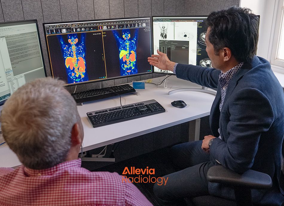
Nuclear medicine is a form of medical imaging or scan that uses small amounts of radioactive tracer substances to produce images of your body. It’s also referred to as nuclear medicine scans, nuclear scans, nuclear imaging or radionuclide imaging.
- Specialised cameras are used to track the path of these radioactive tracers to create computer images of organs and tissues inside your body.
- Nuclear scans provide information on how organs, tissues, and cells are working. Other common imaging procedures, such as X-ray, only show the structures.
The radioactive tracer is also known as a radiotracer, radionuclide, or radiopharmaceutical.
Video: What to expect – nuclear medicine
Nuclear medicine scans are mostly used to diagnose cancer but may also be used for other illnesses. There are several different nuclear scans – the following are a few examples.
- Positron emission tomography (PET) scans: PET scans are usually used to examine your entire body from skull to thighs, although some PET scans can be limited to certain organs, such as your brain or heart only. Different types of radiotracers are used depending on the reason for the scan. Read more about PET scans and how they work.
- Thyroid scans: These check how your thyroid is working, or they may look at a thyroid nodule. A very small amount of radiotracer is injected into one of your veins, which is then taken up by your thyroid. A special camera called a gamma camera is then used to give a picture of the radioactive tracers in your thyroid gland. Read more about thyroid scans(external link).
- Bone scans: These can check for problems in your bones (eg, diseases, tumours, or the cause of pain or inflammation) or the joints. The radioactive tracers collect in your bones over a few hours before the scan starts. Read more about bone scans.
- Radionuclide angiography: This scan uses radioactive tracers to track how well the chambers in your heart are working and how well your heart pumps blood.
Nuclear scans are mostly safe and have few side effects. The radioactive tracers have a low risk of causing an allergic reaction and the doses of radiation are very small. Some people may have pain or swelling at the site where the radioactive tracer is injected into a vein.
References
- Nuclear medicine(external link) National Institute of Biomedical Imaging and Bioengineering, US, 2022
- Facts about nuclear medicine(external link) CDC, US, 2024
- Nuclear medicine(external link) Allevia Radiology, NZ, 2024
Credits: Healthify editorial team. Healthify is brought to you by Health Navigator Charitable Trust
Reviewed by: Dr Remy Lim, Medical Director, Allevia Radiology
Last reviewed: