Ectropion
Key points about ectropion
- Ectropion is when your lower eyelids sag and turn outwards. This can happen in one or both of your eyes.
- The most common reason for is ageing, but some other conditions may also cause ectropion, such as a lump, injury or burn.
- Ectropion isn't usually serious but can be uncomfortable.
- Treatment ranges from eye drops to surgery.
- There are self-care steps you can take to reduce irritation in your eye when you have the condition.

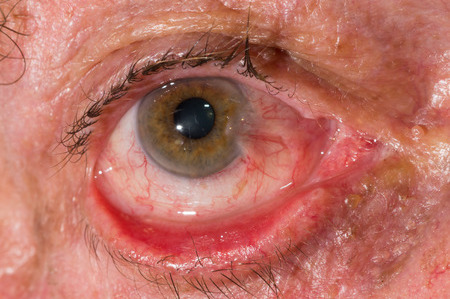
Ectropion is when one or both of your lower eyelids sag and turn outwards. It's usually due to ageing, but some other conditions may also cause ectropion, such as a lump, injury or burn. It's not usually serious but can be uncomfortable.
Treatment of ectropion ranges from eye drops to surgery. There are also things you can do to reduce irritation in your eye, such as using artificial tears and not wearing make-up.

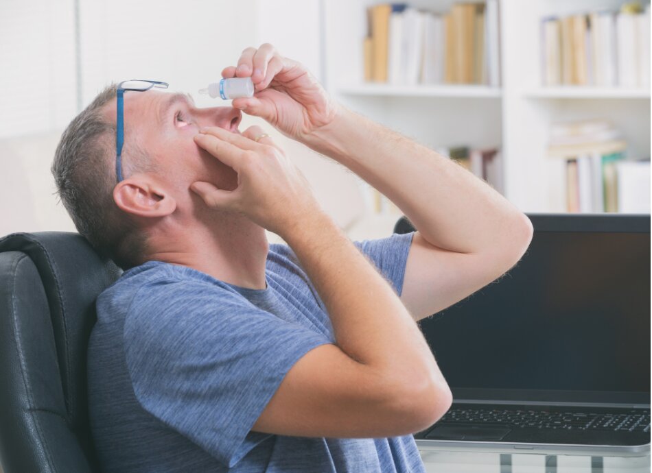
Image credit: Shutterstock
Most commonly ectropion is caused by ageing. The lower lid muscles of your eyes become weaker as you get older and turn outwards.
Other causes of ectropion include having:
- a type of facial paralysis called Bell's palsy
- a lump, cyst or tumour on your eyelid
- a burn
- had previous surgery
- a skin condition called contact dermatitis
- an injury causing damage to the skin around your eyelids.
Common symptoms include:
- excessive tearing
- crusting of the eyelid
- mucous discharge
- irritation of the eye
- conjunctivitis
- corneal ulceration or infection.
You should see a doctor immediately if you have any of these symptoms:
- blurred vision
- loss of side vision
- seeing coloured halos
- redness of your eye
- nausea or vomiting
- pain in your eye.
If you are concerned that you might have ectropion, it is important to see your healthcare provider or optometrist for an eye examination. They are able to detect eye diseases and assess your symptoms. You will then be referred to an eye surgeon (ophthalmologist) for further treatment if necessary. No special tests are needed most of the time.
See your healthcare provider if you have or think you are developing ectropion. Treatment will depend on its severity.
Self-care
While your eyes are irritated, avoid using make-up such as eye shadow, eyeliner and other cosmetics around the eye. Also, avoid using contact lenses until the condition is under control.
If you have dry eyes or mild cases, an eye lubricant such as artificial tears (during the day) or a lubricant ointment (at night) can help to keep your eyes moist.
If your eyes become increasingly red or painful, or your sight becomes blurred, you should seek urgent care from your optometrist or eye surgeon.
Surgery
The surgery for treatment of ectropion is called eyelid surgery and will depend on the reason for your ectropion. Surgical techniques could include lid-tightening procedures, punctal opening (opening of the tear ducts) and the addition of skin with a skin graft. Your eye surgeon or ophthalmologist will be able to recommend the best procedure for you.
Regular eye checks are important
As you get older, you should get your eyes checked regularly. Also get your eyes checked straight away if you notice any changes or think your vision is not as clear as it used to be.
Eye protection
Wear sunglasses to reduce glare and UV damage.
Ectropion(external link)(external link) NHS Choices, UK, 2018
Ectropion(external link)(external link) MedlinePlus, US, 2018
Auckland Eye(external link)(external link) NZ
Ectropion(external link)(external link) Cleveland Clinic, US, 2022
The following information has been adapted from The Auckland Eye Manual(external link).
General description
An ectropion is a positional problem of the eyelid in which the margin is rotated away from the globe. It is generally an involutional change related to laxity of the canthal tendons and lower lid retractor disinsertion. An ectropion can also be cicatricial related to skin shortage, mechanical from lid lumps or paralytic from seventh nerve palsy.
Symptoms
Commonly, patients have irritation from conjunctival thickening and drying. Watering relates to lid margin and punctal displacement along with secondary punctal stenosis.
Signs
There may be an elevated tear film (increased height of tear meniscus along the lower lid margin) and conjunctival inflammation in the region of the ectropion. Laxity of the medial or lateral canthal tendons is commonly seen. In cicatricial ectropion the skin can be generally shortened from sun damage or related to a scar. A paralytic ectropion will have the associated features of a facial palsy.
Slit lamp signs
Punctal stenosis is best seen on the slit lamp and this may also show maceration of the conjunctiva from prolonged exposure. In paralytic ectropion corneal drying, erosions and ulceration can occur from incomplete eyelid closure.
Immediate management
For corneal drying, regular lubricants are used (Lacrilube or Polyvisc ointment at night and artificial tear drops during the day). If there is any corneal ulceration, this must be managed with appropriate topical antibiotics. In cases of acute paralytic ectropion, the lower lid can be taped for support and Botox injected into the levator muscle to produce a temporary tarsorrhaphy.
Long-term management
In general, ectropion is well treated with surgery aimed at rectifying the underlying causes. This may include lid-tightening procedures, punctal opening and the addition of skin.
Referral guidelines
Most ectropions can be referred on a non-urgent basis unless there is concern regarding the health of the cornea.
Credits: Adapted by Healthify editorial team with permission from The Auckland Eye Manual 2021
Reviewed by: Kenny Wu. Optometrist, Eye Institute, Auckland
Last reviewed:
Page last updated:


