Key points about X-ray

  • An X-ray uses a small amount of radiation to create images of your bones and internal organs. 
  • X-rays are most often used to detect bone or joint problems, or to check the heart and lungs.
Looking at X-ray Mercy Radiology
Print this page

X-rays are a form of electromagnetic radiation, similar to visible light. However, X-rays have higher energy than light and can pass through most objects, including the body.

By placing an X-ray detector or plate on the other side of a person, an image will be formed that represents the “shadows” created by the objects inside the body.

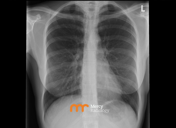
Structures containing:

  • air (such as the lungs) will be black,
  • muscle, fat, and fluid will appear as shades of grey and
  • dense structures such as bones will block most of the X-ray particles, and will appear white.

Video: What is an X-ray?

(Mercy Radiology, NZ, 2019)


You will be asked to remove your watch, jewellery or garments with metal closures from the part of your body being imaged. These items can block part of the image.

  • You may be asked to wear a gown.
  • You may be asked about your overall health or any medications you take.

Let the radiographer (the person who performs your X-ray) know if you:

  • are or may be pregnant
  • have had an X-ray of this part of your body before
  • have metal (eg, a pacemaker or a surgical pin) in the part of your body being imaged.

During your X-ray

  • You will be asked to lie on a table, sit or stand.
  • A lead apron may be draped over part of your body to shield it from the X-rays.
  • With an X-ray of your chest or abdomen, you will have to take a deep breath and hold it for a few seconds.
  • Having an X-ray is like having a photo taken. You need to hold still and you will not feel anything. 
  • While having an X-ray is painless, sometimes the position needed for the best view of the area being X-rayed is uncomfortable for a minute or two.
  • For best results, remain as still as you can during your X-ray exam.
  • These days, having an X-ray is very quick and most only take 5 to 10 minutes. 

After your X-ray

The films or images will be viewed by a radiologist (doctor who specialises in imaging) who will describe what the X-ray shows. This report will then be sent to your doctor who will discuss the test results with you during a follow-up appointment or over the phone.

Some people are concerned that having an X-ray increases your chance of getting cancer. However, it is also believed that you would have to be x-rayed many, many times to receive the amount of radiation that would be bad for your health. The amount of radiation you get from having a chest or limb X-ray is much less than the Earth’s natural radiation you are exposed to every day.

A chest X-ray is a ‘picture’ that shows the ribs, lungs, diaphragm and size of the heart. 

  • When someone is acutely unwell with shortness of breath, fever and cough, a chest X-ray might be ordered to look for signs of infection, (such as pneumonia) inflammation, fluid build up in the lungs or tumours or masses.
  • It is often used to assess people who have smoked for many years to look for signs of chronic lung disease and lung cancer.
  • A chest X-ray is also often done before an operation to check the lungs and heart appear normal.


Chest X-ray supplied by Mercy Radiology

 Image credit: Mercy Radiology

Read more about chest X-ray.

  • If you have ongoing pain in a joint such as a hip, knee or hands, you may have an X-ray to look for signs of arthritis. 
  • Joint or limb X-rays are also done after an injury to look for broken bones or other causes of the pain. 
  • Structures containing air will be black, and muscle, fat, and fluid will appear as shades of grey.

In Aotearoa New Zealand, radiology services such as X-rays, are available through public and private providers. 

Public Services
Find radiology services in your region(external link) HealthPoint NZ

Private Services
Find private radiology services in your region(external link) HealthPoint NZ

X-ray – an introduction(external link) NHS, UK, 2022
X-rays – what are medical x-rays and how do they work?(external link) National Institute of Biomedical Imaging and Bioengineering (NIBIB), 2022
X-rays (plain radiography)(external link) InsideRadiology, The Royal Australian and New Zealand College of Radiologists
X-rays (Radiolography)(external link) RadiologyInfo, US

Apps

Self-management and healthy living apps

Need help now?

Healthline logo in supporters block

Need to talk logo

Healthpoint logo

Credits: Healthify editorial team. Healthify is brought to you by Health Navigator Charitable Trust.

Reviewed by: Dr Janine Bycroft, GP

Last reviewed:

Page last updated: