You can now add Healthify as a preferred source on Google. Click here to see us when you search Google.
Retinal detachment
Key points about retinal detachment
- The retina is the layer of tissue at the back of the eye that detects shape and colour.
- When the retina separates from the inner lining within the eye, it's known as retinal detachment.
- Retinal detachment is an uncommon but serious eye condition which can cause blindness if not treated promptly.
- Most retinas can be reattached with surgery.
- To reduce vision loss, retinal detachment needs to be diagnosed and treated promptly.

How the retina works
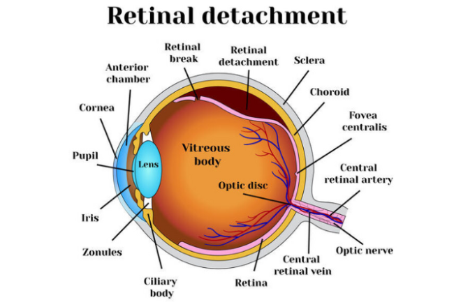
The retina is the innermost wall of the eye. It is made up of light-sensitive cells called rods and cones, which detect shape and colour. Light enters the eye and is focused on the retina by the lens. The retina makes a picture from the light and sends this image to the brain via the optic nerve. If the retina is damaged, the image will be distorted.

Image credit: 123rf
Causes of retinal detachment
Retinal detachment occurs when small holes or tears occur in the retina and allow fluid to seep inside. This creates pressure within the retina which causes the rods and cones to become separated from the underlying tissue. The most common cause of retinal tears is the shrinking of the vitreous (a thick, jelly-like substance within the eyeball that keeps it firm). As the vitreous shrinks, it pulls the retina away from the back of the eye.
Another cause of retinal detachment is scar tissue caused by inflammation. The scar tissue can tug on the retina, causing it to pull away from the underlying tissue. This can be a complication of diabetic eye or other eye diseases. Rarely, fluid can seep out of blood vessels in the eye and cause the retina to be separated.
A retinal detachment can also be caused by an injury or trauma to the eye.
Who is at risk?
There are about 400 cases of retinal detachment per year in New Zealand. In most cases, there is no clear trigger. It is more common in patients with myopia (short sightedness), the elderly and those with a family history of retinal detachment.
Retinal detachments are painless but serious. Symptoms may include:
- flashes of light – these are most obvious in dim lighting and in your side vision
- sudden appearance of 'floaters' – small, moving spots or specks in the field of vision*
- visual disturbance such as shadowing in your side vision or the sensation of a curtain coming over your eye
- blurred, cloudy or distorted vision.
* Floaters are common. If they occur on their own, they are usually not a major cause for concern. As a rule of thumb, let your optometrist know about any new floaters and seek urgent advice if you have new floaters as well as flashes of light.
Contact your GP, optometrist or eye specialist immediately if you get any of these warning signs. The sooner retinal detachment is treated, the better the outcome.
Video: What Are The Symptoms Of Retinal Detachment | Specsavers
This video may take a few moments to load.
(Specsavers, UK, 2016)
Most people with retinal detachment need surgery. If your central vision has not been affected, retinal detachment repair should be carried out as soon as possible. If central vision has already been lost, the timing of surgery is less critical. Surgery involves reattaching the retina on the inside of the eye. This usually involves the use of a laser to 'spot weld' the retina back on. Often a gas bubble is placed inside the eye to help hold the retina steady while it is healing.
If a gas bubble is used, you will need to hold your head in a certain position following surgery. This is so that the bubble is in the right place to support the healing retina. Your surgeon will tell you what position you need to hold your head in and for how long. The gas usually takes about 10 days to resorb and air travel is not possible during this period.
Success of surgery
Around nine out of 10 retinal detachments are successfully repaired with a single operation. However, repairing a detached retina is not always successful. In some eyes, the retina may redetach due to the development of new retinal tears or scarring on the surface of the retina. This may mean you need to have another operation.
Sometimes, vision may remain poor even when the retina is successfully reattached. This is commonly due to irreparable damage to the retinal cells that occurred when the retina detached and before treatment was sought. For this reason, it is vital that medical advice is sought as soon as possible if you have symptoms of retinal detachment.
There are many causes of retinal detachment and the triggers aren't fully understood. Looking after your eyes is the best way to prevent eye damage. Make sure you:
- Use protective eye wear to prevent eye trauma.
- Control your blood sugar carefully if you have diabetes.
- See your eye care specialist once a year (you may need more frequent visits if you have risk factors for retinal detachment).
- Are alert to symptoms of new flashes of light and/or floaters.
Retinal detachment(external link) Medline Plus, US
Retinal detachment (external link) Eye Smart, Eye health information from American Academy of Ophthamology
Credits: Healthify editorial team. Healthify is brought to you by Health Navigator Charitable Trust.
Reviewed by: Karen Moulton, Optometrist, Bay of Plenty
Last reviewed:
Page last updated:


