Fluoroscopy
Key points about fluoroscopy
- Fluoroscopy is a type of X-ray procedure that shows your internal organs and parts of your body moving in real-time.
- It's used to diagnose certain conditions and to provide visual guidance to healthcare providers during procedures.
- Because fluoroscopy uses X-ray, it's important to inform your healthcare provider before any fluoroscopic procedure if you're pregnant.
Fluoroscopy is a medical imaging technique that uses low dose X-ray to take a series of continuous images of the relevant body parts. If an X-ray is like a photograph, fluoroscopy is like a short video.
Healthcare providers use fluoroscopy for 2 main reasons:
- To see inside your body and diagnose (identify) certain conditions.
- To visually guide them during procedures, eg, surgery, placement of stents or catheter.
Diagnostic fluoroscopy
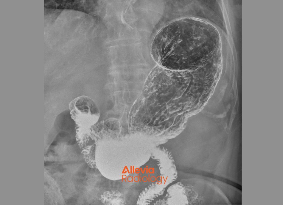
Barium swallow – done to check for problems in your throat, oesophagus, stomach and parts of your small intestine. You swallow a drink containing barium (known as a ‘contrast’) which shows up on the fluoroscopy images. Read more about barium swallow and barium enema (occasionally done to look at the lower part of your bowel).
The image below is of a barium swallow.
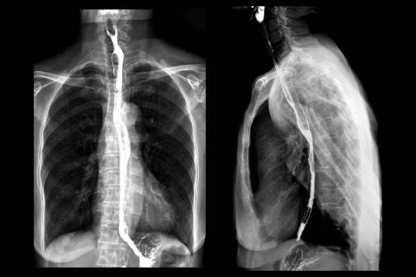
Image credit: Canva
Gastrografin enema – a fluoroscopy study of your large bowel (colon). Gastrografin is a colourless liquid contrast that would be used to highlight your large bowel.
Angiography – a procedure using contrast injected into a blood vessel. It shows blood flow through the arteries and veins of your body to find any blockages, narrowing or aneurisms (bulges in the wall of a blood vessel). Read more about angiography.
The image below is an angiogram of the heart blood vessels.
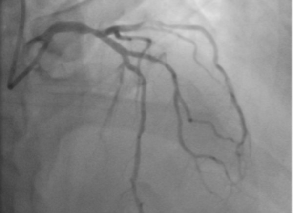
Image credit: Allevia Radiology
Cystogram – a procedure using fluoroscopy imaging to help diagnose problems in your bladder.
Urethrogram – Fluoroscopy imaging of the urethra (the tube from your bladder to the outside) almost always in men.
Discogram – an imaging test to look at discs of your spine and sometimes for the cause of back pain. This procedure can also be done using CT scan.
Myelogram – an imaging test to get detailed pictures of the structures of your spine and spinal cord.
Arthrogram – a procedure to look at a joint, such as your shoulder, hip or knee. Often a dye or contrast will need to be injected into the joint to show more details for a proper diagnosis. These are often done together with an MRI study.
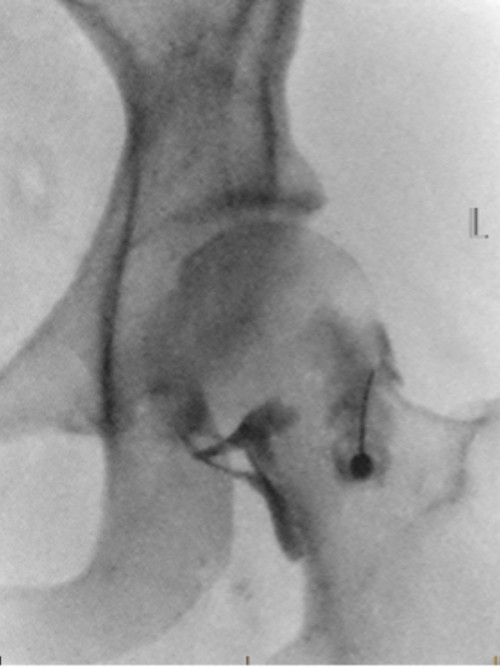
Image credit: Allevia Radiology
Fluoroscopy guided procedures
Angioplasty – placement of stents to widen blocked or narrowed arteries to improve blood flow. Commonly performed on the blood vessels of the heart. Read more about angioplasty.
Orthopaedic surgery – fluoroscopy might be used by your surgeon to help guide procedures such as joint replacement or fixing broken bones.
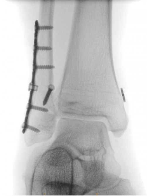
Image credit: Allevia Radiology
Catheter insertion – fluoroscopy can help with the proper placement of catheters to help get fluids into or draining from your body.
Joint injections – fluoroscopy can be used to help healthcare providers inject steroids or anaesthetic into a joint such as shoulder or hip.
Any preparation required depends on the type of procedure and why you're having it. Some tests don't need any special prep. When you’re booking your appointment, you’ll be told if your test requires anything specific, eg, if you should fast (not eat or drink) for several hours beforehand.
If you're pregnant, or think you might be, it’s important to let your healthcare provider know.
Also, tell your healthcare provider if you have any allergies or have had a reaction to contrast dye before.
Your fluoroscopy might include these steps:
- Removing clothing and jewellery that might interfere with the procedure. Don't worry, you’ll be given a gown to wear.
- Contrast dye might be used to make a part of your body show up on X-ray. It might be a liquid you drink, or it may be injected into your joints, veins or arteries, or the part of your body that's being imaged.
- Usually you'll be asked to lie on an X-ray table and you might be asked to move to, or hold, a certain position.
- The doctors and imaging technologists will use the fluoroscopy machine to watch and guide what they're doing on the images produced.
Depending on what procedure you're having, you might be seen as an outpatient and be able to go home afterwards. Sometimes you'll need to stay in hospital following the procedure, eg, for angioplasty.
What you experience after your fluoroscopy procedure will depend on which procedure was done, eg:
- The barium drink used in a barium swallow study can sometimes cause constipation. Increasing your fluid intake for a few days after the procedure is recommended. Read more about constipation.
- If you’ve had a discogram or a myelogram you may experience back pain for several hours.
- In the 24 hours after a myelogram you may experience headache, nausea (feeling sick) or vomiting (being sick). These side effects should pass.
- Any expected post procedure effects will be explained by the staff during your procedure.
Generally, the amount of X-ray you receive from a fluoroscopy procedure is very small. However, your exposure to radiation will increase depending on the complexity of your procedure.
The benefits of the fluoroscopic procedure usually outweigh the risks.
Remember, if you’re pregnant or think you might be, please talk to your healthcare provider first to see if a procedure involving the use of radiation is suitable.
References
- Fluoroscopy(external link) Allevia Radiology, NZ
- Fluoroscopy(external link) Cleveland Clinic, US, 2021
- How fluoroscopy works(external link) How Radiology Works, US
Credits: Healthify editorial team. Healthify is brought to you by Health Navigator Charitable Trust.
Reviewed by: Peter Hsu and Penny Marks, Medical Imaging Technologists, Allevia Radiology
Last reviewed: