Macular holes

Key points about macular holes

  • A macular hole is a small puncture in the centre of the macula area of the retina.
  • Macular holes are more common in people over the age of 50.
  • At first, you may notice distortion or blurring if you have a macular hole. The only way to find out is to get an eye examination.
  • Without treatment, a macular hole can lead to complete loss of central vision in the affected eye.
  • Treatment for macular holes is eye surgery called vitrectomy, which aims to seal the hole.
  • Treatment works well for most, but not all people.


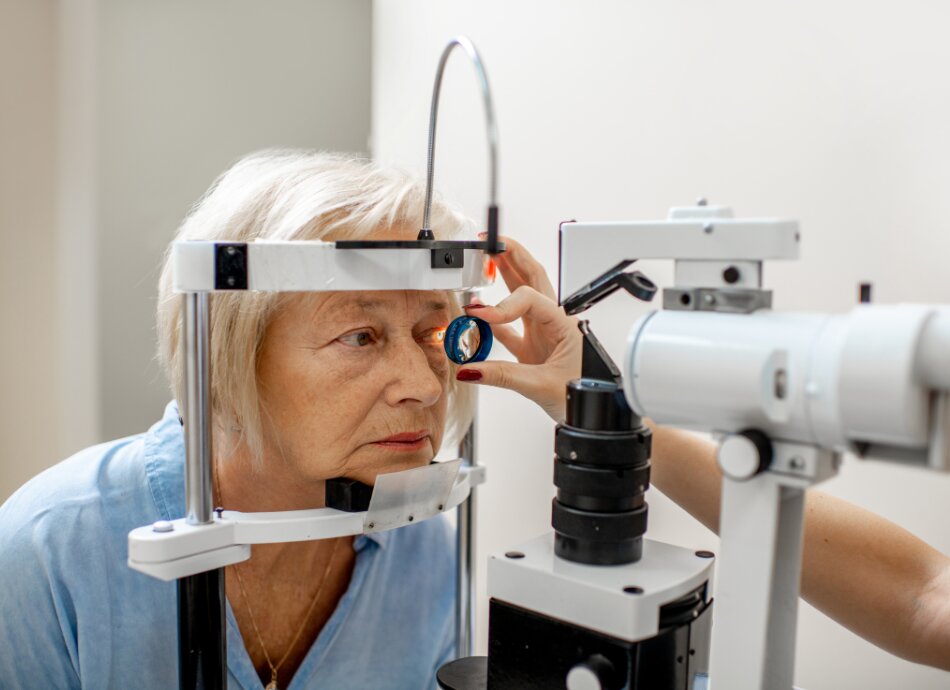
Older woman leaning forward to have eye examination
Print this page

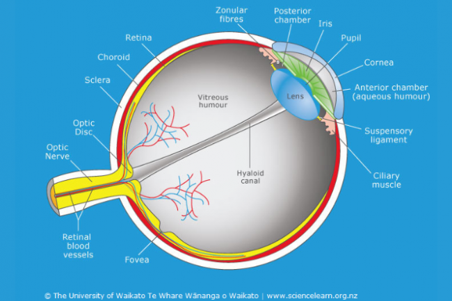
The retina is a clear film of very delicate tissue that lines the inside of the back of the eye. It senses light that shines into the eye and send signals through the optic nerve to the brain. The brain interprets these signals as visual images.

Image credit: Science Learning Hub – Pokapū Akoranga Pūtaiao, University of Waikato, www.sciencelearn.org.nz

The macula is the central area of the retina that gives us sharp central vision and reading vision. The very central portion of the macula, the fovea, is the thinnest and most delicate portion of the entire retina. It is in this very thin, delicate fovea area that a macular hole can develop.

A macular hole and macular degeneration are completely different conditions affecting the retina. However, the potential outcome of loss of central vision in the affected eye is common to both conditions.

In most cases, a macular hole develops because of normal physical changes that occur within the eye. These changes are not due to anything the person has done. They are natural changes that only cause problems in some people.

These changes put stress on the macula and may cause a hole to form in the most delicate area, the fovea. The hole expands just like a tear in a nylon stocking might enlarge. As it enlarges, fluid passes under the retina, which in turn causes the hole to get larger. This type of macular hole occurs most commonly in people over 50 years of age and is commonly called an ‘idiopathic’ macular hole.

Other rarer causes of macular holes include:

  • severe blunt injury to the eye
  • retinal detachment
  • conditions that cause severe swelling of the retina.

In the early stages, the macular hole is very small, so the central vision may be only slightly blurred or distorted. As the hole enlarges over several weeks to several months, the symptoms progress. The hole can enlarge to a point at which the affected eye can only see the larger letters of an eyesight testing chart.

A macular hole can't make your eye go completely blind. It affects only the very centre of your vision and does not cause a loss of the peripheral (side) vision.

If a macular hole is suspected, you may need to have an optical coherence tomography (OCT). This is a simple procedure that creates an image of the retina at the back of the eye. This imaging, which is similar to a CT (computed tomography) scan used for other parts of the body, can show the hole in the macular.

A macular hole can be treated and repaired with surgery called a vitrectomy. The surgery is usually done as a day (outpatient) surgery and there is a greater than 95% success rate. There is no other treatment for a macular hole.

During a vitrectomy:

  • You will be given medicine to help you relax and your eye will be numbed to dull the pain (anaesthesia).
  • The surgeon will make very small holes through the white part of the eye.
  • These holes are so small that usually no stitches are required.
  • Using a microscope, the surgeon can use a variety of very specialised instruments placed through these holes to work within the eye.
  • The gel-like substance that fills the eye is removed and replaced with a specially designed saline solution.
  • The surgeon then peels a very thin membrane from the surface of the macula surrounding the macular hole.
  • Peeling this membrane is important for helping the macular hole to seal.
  • Finally, a gas bubble that completely fills the back of the eye is used to replace the saline solution.
  • The gas bubble will gradually go away after surgery and is replaced by the gel-like fluid that is produced normally inside the eye.
  • Laser and freezing treatment may also be used to secure the retina in place.

Care after surgery

  • A patch is worn over the eye until the morning after surgery.
  • Eye drops that help healing are used several times each day for four weeks after surgery.
  • People are usually asked to position themselves face down for five days immediately following the operation. Positioning face down allows the gas bubble to press firmly against the macular hole and may slightly increase the chance of the hole closing well. Your eye specialist will discuss this with you.

Outcomes of surgery

Usually, vision recovers to ‘normal’ (6/6) or to the vision required for a driver's licence (6/12) after successful macular hole surgery. However, some people may have more limited improvement in vision and a small percentage of people may not improve very much at all even after successful surgery. 

The amount of visual improvement depends on:

  • whether the macular hole closes
  • the person's age
  • the physical condition of the macular hole
  • other existing eye problems.

After the surgery it can take anywhere from 3–18 months for your vision in the affected eye to reach its best.

Possible complications of macular hole surgery

Any surgical procedure carries a risk of complications and macular hole surgery is no exception. There are 3 main complications of macular hole surgery to be aware of:

Infection of the eye after surgery: Most infections can be effectively treated if picked up at an early stage. However, there is a risk that an infection can create severe damage that could lead to blindness in the affected eye. Fortunately, eye infections after surgery are rare, occurring in only 1 of 2000 cases.

Retinal detachment: This can occur at any time, even if there has been no surgery. However, if you have had macular hole surgery, your eye is at greater risk of developing retinal detachment. A retinal detachment can occur soon after surgery, and occasionally it can develop months or years later. It can lead to blindness if not repaired. Fortunately, nearly all retinal detachments can be repaired with additional surgery. Retinal detachment after macular hole surgery occurs for 1 or 2 out of every 100 people. Ongoing improvements in surgical techniques and instruments are reducing this likelihood.

Cataracts: These cause haziness in the lens of the eye and commonly develop as a natural consequence of ageing. Having a vitrectomy causes cataracts to progress more quickly. Cataract surgery may be needed within 1 year of vitrectomy surgery.
For further information talk to an optometrist or eye specialist. Optometrists are listed in the Yellow Pages of your telephone book. Eye specialists are listed with registered medical practitioners at the front of the White Pages of your telephone book. You can use this link to search for an optometrist near you – type optometrist into the search bar. 

Macular hole(external link) Eye Institute Auckland, NZ

Need help now?

Healthline logo in supporters block

Need to talk logo

Healthpoint logo

Credits: Kenny Wu, Optometrist, Eye Institute, Auckland

Reviewed by: Healthify editorial team. Healthify is brought to you by Health Navigator Charitable Trust.

Last reviewed:

Page last updated: